asthma x ray findings|Radiographic characteristics of asthma : Cebu Asthma is one of the most common chronic diseases in the world. According to the 2014 Global Asthma Report, it is estimated that around 300 . Tingnan ang higit pa Check out our complete 1x2 betting explained guide to learn what it is, how it works, how to bet on 1x2 markets, its pros and cons, and tips for successful bets. Betting sites. by region. Europe. UK; . If you don’t understand what a lay bet means, it is when bettors place a wager against an outcome occurring. Instead of betting on another .

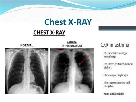
asthma x ray findings,Plain chest radiographscan be normal in up to 75% of patients with asthma. Reported features of asthma include: 1. pulmonary hyperinflation 2. bronchial wall thickening: peribronchial cuffing (non-specific finding but may be present in ~48% of cases with asthma 1) 3. pulmonary edema (rare): . Tingnan ang higit pa
Asthma is one of the most common chronic diseases in the world. According to the 2014 Global Asthma Report, it is estimated that around 300 . Tingnan ang higit paThe classical symptoms of asthma are wheeze, shortness of breath, chest tightness or difficulty breathing and cough. These symptoms are typically . Tingnan ang higit paInflammation plays a major role in asthma and involves multiple cell types and mediators. The factors that initiate the inflammatory process are complex and still under investigation. Genetic factors (such as cytokine response profiles) and environmental exposures (such as allergens, pollution, infections, microbes, stress) at a crucial . Tingnan ang higit paThe goal of the treatment is to control the symptoms, prevent exacerbations and loss of lung function and reduce associated mortality. Drugs used to control asthma depend on the severity of the disease. Short-acting β2-agonists can be used in patients with mild occasional symptoms. Inhaled steroids (oral steroids might be required . Tingnan ang higit pa In acute asthma, a chest x-ray is only required if there is 4: suspected pneumomediastinum, pneumothorax or surgical emphysema; suspected consolidation; requirement for ventilation or .
Chest radiography is the initial imaging evaluation in most individuals with symptoms of asthma. The value of chest radiography is in revealing complications or alternative causes of wheezing.They found that compared to controls, those with severe asthma have 2.3 times higher median ventilation defect percentage and that ventilation defects decrease over time .The clinical aspects of asthma are paroxysmal respiratory distress, recurrent cough, wheezing, and chest tightness. However, the radiographic traits of asthma have seldom .
A chest X-ray can help identify any structural abnormalities or diseases (such as infection) that can cause or aggravate breathing problems. Allergy testing. Allergy .
Healthcare professionals conduct a variety of diagnostic tests for asthma. Learn about the use of chest X-rays and other tests to diagnose asthma. X-rays send small amounts of electromagnetic radiation through your chest to create images of the bones and tissues. In terms of an asthma diagnosis, an X-ray of . Asthma is a common disorder characterized by chronic airway inflammation, airway hyperresponsiveness, and variable airway obstruction that affects >300 million .Asthma is one of the most common diseases of the lung. Asthma manifests with common, although often subjective and nonspecific, imaging features at radiography and HRCT. Perhaps of utmost .
In children with asthma, routine chest X-ray typically shows bilaterally increased air volume, low diaphragms, wide diaphragmatic angles, and often a slender cardiac silhouette with a prominent pulmonic arch. Such an X-ray is not diagnostic of asthma itself, however, but rather of its complications: pneumonitis (particularly in toddlers with .asthma x ray findings Radiographic characteristics of asthma Typical clinical findings in asthma may include: Around the bedside: oxygen, inhaler and spacer, PEFR meter; . A chest X-ray is also important to rule out infection, collapse or pneumothorax. Other. .
Pulmonary findings in Churg–Strauss syndrome in chest X-rays and high resolution computed tomography at the time of initial diagnosis July 12, 2010 | Clinical Rheumatology, Vol. 29, No. 10 Imaging of Asthma .Radiologic Findings of Bronchial Asthma Jai Soung Park, M.D., Sang Hyun Paik, M.D. Department of Radiology, Soonchunhyang University School of Medicine Asthma is the most common disease of the lungs, and one that po ses specific challenges for the physicians including radiologist. This article reviews for the clinical diagnosis, R adiologic .A chest X-ray can help accurately diagnose a patient’s condition that may be affecting breathing. Here at Independent Imaging, you can trust our experts to provide you with the best imaging services for your diagnostic needs. Our highly qualified radiologists are ready to assist you. To make an appointment, call us at (561) 795-5558.Also known as a chest X-ray, a chest radiograph uses an X-ray beam. It is considered the best general technique for looking at the lungs, the chest cavity, and the area around the lungs. 1,2. To undergo a chest X-ray, you will typically stand in front of a wall-mounted device that holds the X-ray film or a plate that digitally records the image. Chest X-rays expose the patient briefly to a minimum amount of radiation. Any radiation exposure has some risk to the tissues of the body. The radiation . In a study by Lo et al of 30 children with difficult-to-treat asthma, abnormal CT findings were highly prevalent in a cohort of children with severe asthma, with bronchiectasis identified . Imaging Findings. Asthma typically involves mainly small and medium-sized bronchi. Air-trapping on expiratory scans most common finding. Bronchial wall thickening (50-90%) Decreased lung attenuation (50%) Mosaic lung attenuation. Degree of mosaic attenuation correlates with degree of asthma. HRCT useful to evaluate for bronchiectasis.

High-resolution CT manifestations of asthma include thickening of the bronchial wall, narrowing of the bronchial lumen, areas of decreased attenuation and vascularity on inspiratory CT scans, and air trapping on expiratory CT scans [ 1 – 3 ]. Other findings that are seen with increased frequency in patients with asthma are bronchiectasis and .Radiographic characteristics of asthma High-resolution CT manifestations of asthma include thickening of the bronchial wall, narrowing of the bronchial lumen, areas of decreased attenuation and vascularity on inspiratory CT scans, and air trapping on expiratory CT scans [ 1 – 3 ]. Other findings that are seen with increased frequency in patients with asthma are bronchiectasis and . Physical exam. Your healthcare professional may: Examine your nose, throat and upper airways. Use a stethoscope to listen to your breathing. Wheezing — high-pitched whistling sounds when you breathe out — is one of the main signs of asthma. Examine your skin for signs of allergic conditions such as eczema and hives. CT stands for computed tomography. It uses X-rays to build a 3-dimensional picture of the inside of your body. This gives a detailed picture of your lungs, blood vessels and other organs. You may be given an injection of material that shows up on the scan and can help to outline blood vessels. This is called contrast.

Chest x-rays at diagnosis should be reserved for children with severe disease or in any patient with atypical features or clinical symptoms or signs suggesting other conditions. . In the absence of .The clinical aspects of asthma are paroxysmal respiratory distress, recurrent cough, wheezing, and chest tightness. However, the radiographic traits of asthma have seldom been reported and found. Park reported that acute asthma reversibly increases lung compliance and total lung capacity (TLC). [ 1] The authors confirm anecdotal evidence of .
Predilection. Siamese cats are predisposed to lower airway disease, with a breed prevalence of up to 5%.4 Some authors suggest there is no sex predilection, 1 while others have documented that females are affected more significantly. 3 Asthma develops in young to middle-aged cats, with a reported mean age of 4 years (range, 1–15 years). 1 . Learn about the latest asthma x-ray findings and how Nao Medical's pulmonology specialists can help you manage your asthma. Skip to content Nao Medical After Hours service is currently available!
asthma x ray findings A chest X-ray can help identify any structural abnormalities or diseases (such as infection) that can cause or aggravate breathing problems. Allergy testing. . Long-term asthma control medications, generally taken daily, are the cornerstone of asthma treatment. These medications keep asthma under control on a day-to-day basis and .In asthmatic patients CXR can show nonspecific findings, including peribronchial thickening and hyperinflation. Its primary utility is to exclude diagnoses with similar symptoms or to evaluate for complications, including infiltrates, pneumothorax, or pneumomediastinum. Studies have shown that CXR results rarely change clinical .The National Heart and Lung Institute's guidelines advise against routine use of chest X-ray (CXR) for acute asthma exacerbation. Despite this recommendation, unnecessary CXR are still being performed. The objective of this study is to assess the association between abnormal CXR findings and the severity of asthma exacerbation and its impact on .
asthma x ray findings|Radiographic characteristics of asthma
PH0 · The emerging role of quantitative imaging in asthma
PH1 · The Role of Imaging in the Assessment of Severe Asthma
PH2 · Radiographic characteristics of asthma
PH3 · Imaging of Asthma
PH4 · Chest X
PH5 · Can you detect asthma on an X
PH6 · Asthma Imaging and Diagnosis
PH7 · Asthma (summary)
PH8 · Asthma